Dogs and Knee Surgery: Understanding TPLO Surgery
Dogs rupturing their cranial and caudal cruciate ligament (CCL) causes lameness in their rear legs, often requiring surgery.
Dogs have a cranial and caudal cruciate ligament, like a human’s anterior (ACL) and posterior ligaments. The cranial cruciate ligament (CCL) is the one most commonly torn and is one of the major stabilizers in the knee. Rupturing the CCL is one of the most common causes of lameness in the rear legs of dogs. Veterinarians often perform tibial plateau leveling osteotomy (TPLO) surgery to fix the tear.
Are their risk factors for cruciate tears?
Almost all breeds of dogs and cats are at risk, but some breeds are predisposed to a CCL tear. It is seen most often in Labs, Golden Retrievers, German Shepherds, Boxers, Pit Bulls, American Bulldogs, Mastiffs, Rottweilers, and Newfoundlands.
Poor physical condition and obesity predispose dogs to tearing the ligament, but fit dogs can also get a tear. Ligament tears in dogs are different than in humans. In dogs, the ligament starts to degrade and becomes weak over time whereas humans usually tear during sports. Since one ligament degrades and fails, 40-60% of dogs tear the other knee within two years of the first injury.
The average age in dogs is 2-5 years, but our surgeons have seen dogs as young as 5 months or as old as 14 years.
What are the signs of CCL tears?
Dogs can present in a variety of ways. Some dogs will suddenly become lame on the leg and may hold it in the air. Some dogs will have a mild lameness for months that will get better and worse over time. Other dogs will have the intermittent mild lameness and then suddenly become very lame.
However, if both knees are torn at the same time (which occurs in 30% of dogs), they can present unable to walk in the rear legs. Other signs of dogs with bilateral CCL tears can be difficulty going up stairs or jumping into the car, and difficulty sitting down and placing both legs out to the side (rather than sitting square).
How is a CCL tear diagnosed?
Physical examination is the most common method of diagnosis. The knee will feel thickened from the bones shifting back and forth since there is no ligament holding them together. They will have joint swelling, pain on hyperextension of the knee, and will often sit with the leg kicked out to the side.
The tibial thrust test and the cranial drawer test are the two main tests for instability in the knee.
- For the tibial thrust test, the dog often stands (it is less stressful) and your veterinarian will hold the femur (thigh bone) stable while bending the foot. The tibia (shin bone) should not move, but if the cruciate is torn, then it will thrust forward. This happens because the top of the tibia is on a slope and the cruciate holds the femur on the slope. I make an analogy of a wagon being held on a hill by a rope. If the rope is cut, the wagon will roll down the hill. If the cruciate is torn, the femur will fall down the hill with every step they take (often crushing and tearing the meniscus (cartilage cushion in the joint).
- The cranial drawer test involves holding the femur and the tibia and trying to move the tibia forwards. But in large strong dogs, it is very difficult to get cranial drawer without sedation.
Your veterinarian may also do an X-ray. Although you cannot see the ligament on an X-ray, there are classic changes seen on an X-ray such as swelling and arthritis. Dogs do not develop arthritis in the knee for no reason, and the most common cause is cruciate tears.

What are the surgical options for CCL tears?
There is no best surgery for cruciate tears, but most surgeons (and veterinary journals and research studies) agree that the TPLO is the best surgery for dogs. TPLO stands for tibial plateau leveling osteotomy. Another potential surgery is the lateral suture.
TPLO Surgery
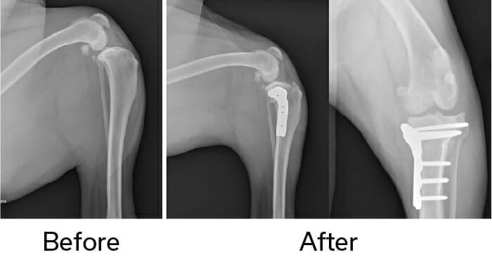
Back to the wagon analogy for the best explanation. If we take the wagon and place it on a flat road, we don’t need a rope to hold it stable. With the TPLO, the surgeon cuts the back part of the tibia with a special circular saw and rotates the piece of bone so the top of the tibia is almost flat. Then the surgeon repairs the bone with a special bone plate to hold it in place while it heals. Once the bone is healed in the “flat road” position, the tibia no longer thrusts forward when the dog walks and the knee is stabilized. The plate usually remains on the bone indefinitely, but it is no longer needed for support once the bone heals.
Lateral Suture
This surgery involves placing a suture, which is usually strong nylon (similar to a strong fishing line), around the outside of the joint to hold the two bones together and prevent them from shifting. The problem with this surgery in large dogs is that there is a lot of force on the nylon and it tends to break and fail. Studies have also shown dogs are in more pain after a lateral suture and develop worse long-term arthritis. Many veterinarians reserve the lateral suture for small breed dogs.

Is TPLO surgery painful?
Using a saw to make a bone cut is different than a trauma causing the bone to break. There is usually not a great deal of swelling or bruising. The TPLO surgery is usually more comfortable than the lateral suture which feels very tight around the joint. Most dogs are using the leg within a few days post operatively, and they will often be more athletic after a TPLO than a lateral suture. Pain management is also critical with using epidurals, infusing the incision with a numbing agent that lasts for three days, and post-operative pain killers.
Can both knees be operated on at once?
Studies have shown similar complications and success of performing bilateral TPLO versus unilateral TPLO. If dogs are under 80-90 pounds, depending on their physique, bilateral TPLO can be performed. In obese or heavier dogs, it is safer to perform one knee at a time. The benefits of a bilateral TPLO are a shorter recovery of six weeks as opposed to 12 weeks. The first week of recovery requires a significant amount of nursing care and support, but usually they are walking quite well by two weeks.
What is the long-term success with the TPLO?
Most data show that 90-95% of dogs will return to their previous activity after the TPLO. All dogs develop arthritis within a few weeks of the ligament tearing due to inflammation, but once the knee is stabilized, this minimizes the arthritis. Dogs will improve up to six months after the surgery as that is how long it takes for them to build muscle strength and for the swelling to improve in the joint. Even with the arthritis, most dogs will only have occasional stiff days after strenuous play or a long rest, but quickly work out of it. Our surgeons have seen dogs going back to work on the Police force and performing agility after a TPLO surgery. However, it does take rehabilitation and muscle strengthening to achieve the best results.
What are the most common complications of a TPLO?
The two most common complications are infection and implant failure/fracture of the bone. Infection is the most common with studies showing an infection rate from 8-17%. The most common reason for infection is that dogs can lick the incision because the E-collar is removed.
If they lick the incision and an infection forms, it can move to cover the plate as there is not a lot of muscle or tissue over the plate. When that happens, it causes a slime coating over the plate that antibiotics cannot penetrate and kill permanently. If an infection forms, the bone usually heals fine, but the implant is often removed to resolve the infection. Fracture/failure is possible, but this is less common with the new Locking Plates and Screws and if the dog is kept restricted and there is no jumping or strenuous play.
How does my dog recover after TPLO surgery?
You should confine your dog to controlled leash walks for six to eight weeks post operatively while the bone heals. They are allowed to walk on the leg, but no jumping, furniture climbing, or running. They should be confined to a crate or very small room while they are healing. We recommend you keep them on a leash so they can be controlled when they are out of the crate, but still feel a part of the family.
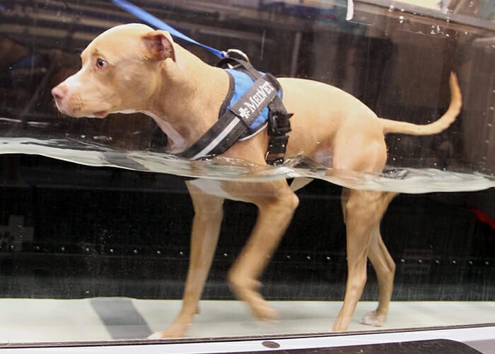
Rehabilitation is recommended to build muscle strength and decrease swelling. Ideally, they will have professional rehabilitation post-operatively, but some clients will choose to perform rehabilitation at home.

With time and a little work, your pet can maintain a good quality of life after TPLO surgery. If you’re concerned about a CCL tear, talk with your family veterinarian about a referral to a MedVet specialist to get your pet on the road to recovery.
FAQs
What is a CCL tear in dogs?
What are the signs of CCL tears in dogs?
How are CCL tears in dogs treated?
Contents


